Reading the Report, Not Just the Result: Interpreting a Private Venous Ultrasound in the UK

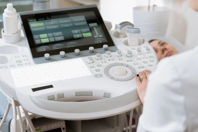
Photo: clinician reading ultrasound scan report at desk, via m.media-amazon.com
A venous duplex ultrasound report is a technical record. It describes what one operator saw, on one machine, with one set of settings, during one appointment of perhaps thirty minutes. It is not a verdict on your health, and it does not establish a diagnosis. Yet because private scans can be booked directly and results often arrive by email within a week, the report is frequently read as though it were conclusive — something to act upon immediately, and often something to pay for again.
CCSVI UK regularly hears from people who have received such a report and are unsure what it means. This article does not assess any individual's results. It explains the structure and vocabulary of these reports, and identifies where interpretation ends and clinical judgement must begin.
What the document contains, and what it leaves out
A typical report has three layers: a description of technique, a set of measurements, and an impression or conclusion. Most patients read the third layer and skip the first two. Clinicians tend to read in the opposite direction, because the impression is only as reliable as the technique that produced it.
The technique section is where the useful detail usually sits. It should say whether you were scanned lying flat, sitting, or both; whether the examination was timed to breathing or to a breath-hold; whether the operator knew your medical history; which veins were examined and at how many points; and what thresholds were applied. If those details are absent, the impression is difficult for anyone — including a second clinician — to weigh.
Reflux, stenosis and flow volume: three words that carry too much weight
Reflux describes flow travelling in the reverse of its expected direction for a measured period. Reports often give a duration in seconds and compare it against a threshold. What the word does not tell you is why the flow reversed, whether it does so consistently, or whether it has any bearing on your symptoms. Reflux is an observation about movement, not an explanation of illness.
Stenosis describes a narrowing. On ultrasound this is usually inferred rather than measured directly — from blood velocity ratios, from an estimate of cross-sectional area, or from the appearance of a valve, septum or membrane. Ultrasound estimates geometry; it does not measure pressure. A narrowed segment may carry entirely adequate flow, and a segment that looks unremarkable may still behave unusually under physiological stress.
Flow volume is calculated from vessel diameter and velocity rather than measured directly. Because cross-sectional area changes with the square of the diameter, even small errors in measurement produce wide margins of error in the reported volume. Figures should be read as estimates with a range, not as fixed quantities.
Why two scans can disagree
Venous imaging is operator-dependent. The angle of the probe, the pressure applied, room temperature, hydration, time of day, posture and the phase of respiration all influence what is recorded. Research studies in this field have typically used fixed protocols and blinded analysis — the person measuring did not know whose veins they were assessing. A routine private examination may do neither.
This is not an accusation of poor practice. It is a statement about method. When the protocol is not described, the reader cannot know how closely the scan resembled the conditions under which published findings were generated, and comparison becomes speculative.
Why findings require clinical context
Venous appearances vary between individuals, and studies have differed on how often apparently abnormal flow patterns are found in people without neurological conditions. That disagreement is precisely why a single measurement cannot settle a question about cause. Nor does any ultrasound finding reliably predict who would or would not respond to an interventional procedure; randomised trial evidence to date has not demonstrated the benefit that early enthusiasm anticipated.
A report describes anatomy and flow. Symptoms are a separate dataset, gathered through history, examination and time. Good clinical reasoning holds the two together; a report read alone holds only one.
A checklist before you act on the findings
- Who performed the scan, and what is their accredited experience in venous duplex work?
- Is the protocol described in enough detail for another specialist to repeat it?
- Are the numerical thresholds used to define reflux or stenosis stated explicitly?
- Are measurements given per side and per segment, with the range of normal made clear?
- Does the report separate observation from interpretation, or present the two as one?
- Was the operator aware of your diagnosis, and were images analysed blinded?
- Will the report be sent to your GP or neurologist, and in what format?
- What would change in your care if the finding were absent?
- What follow-up is recommended, who is accountable for it, and who pays?
What no report can tell you
It cannot tell you why you feel as you do. It cannot tell you whether a procedure would help, or how you would respond. It cannot tell you whether the same examination repeated next month would produce the same numbers. Treating it as though it can is where unnecessary expense and, occasionally, unnecessary risk begin.
Keeping the document in proportion
A report is worth having filed in your NHS records, and worth discussing with a clinician who knows your history. It is also worth a second read from a vascular scientist or neurologist who has no financial interest in what happens next. What it is not worth is a chain of further private scans and consultations undertaken to resolve ambiguity that the first report never claimed to settle.
This article provides general information and does not constitute medical advice. Decisions about investigation or treatment should be made with a clinician who knows your individual circumstances.